
Diagnosis and Testing
Diagnosis
There is not a formal diagnostic procedure for determining if you have a pectus condition. For the most-part a diagnosis can be made just by examining the appearance of the chest. It is typically quite obvious if someone has a pectus condition, just by nature of their sternum dipping in, sticking out, or being symmetrical in shape.
Testing
Most of the time, thoracic surgeons and consultants can broadly classify how severe a pectus deformity is by examining the appearance of your chest. But sometimes, further testing is needed to calculate the severity of a pectus deformity.
Aside from medical testing, it’s also very important that surgeons and consultants speak to patients, to get an understanding of how their life is impacted by the shape of their sternum.
Pectus Carinatum
For patients with pectus carinatum, there are a couple of ways to determine how severe a chest protrusion is. Not all patients with pectus carinatum will undergo testing, as treatment is typically offered based on a visual assessment and examination of the sternum.
Pectus Index (PI)
The Pectus Index is worked out by measuring and comparing the width and depth of the chest, as seen in a CT scan. To calculate the Pectus Index, you divide the width and depth by one another. Below is the most common severity scale used to work out how severe pectus carinatum is:
1.5 - 2.0 : Normal chest with no significant deformity
1.29 - 1.49 : Mild pectus carinatum
1.18 - 1.28 : Moderate pectus carinatum
1.07 - 1.17 : Severe pectus carinatum
Less than 1.02 : Extreme deformity
A consultant might choose to perform a CT scan and calculate the Pectus Index, to help them decide whether a patient would be a better candidate for a chest brace or pectus surgery.
Pectus Excavatum, Arcuatum and mixed deformities
Where the sternum and/or ribcage sinks in towards the heart and lungs, testing is often needed to work out how much a patient is physically impacted by the shape of their sternum. Testing is required in most UK nations, to work out if patients meet the eligibility criteria for surgery on the NHS (or HSC in Northern Ireland).
1: Haller Index (HI)
The Haller Index is worked out by comparing the measurements of the width and depth of the interior of the chest cavity. These measurements are taken from a CT scan of the patient, both on inspiration (whilst breathing in) and on expiration (whilst breathing out). The Haller Index is calculated by dividing the internal chest width by the internal chest depth, which can then be measured on the severity scale below:
Less than 2 = a normal chest
Between 2 and 3.2 = mild pectus excavatum
Between 3.2 and 3.5 = moderate excavatum
Above 3.5 = severe pectus excavatum
Whilst these figures are a good guideline, do not be shocked if you find that your Haller index measures off the scale. In patients with very severe pectus excavatum, the Haller index can reach above 20 and 30.
The Haller Index is a useful way of measuring the severity of pectus excavatum, but it isn’t always reflective of how the shape of a patient’s sternum is affecting their heart and lung function.
2: Data about heart and lung function
If you have a concave chest, a thoracic surgeon or consultant might want to perform tests to see if your sternum is pressing on your heart or lungs, and affecting their ability to function. There are a few different tests that you might have to work out your cardiac and respiratory function:
Echocardiogram
Cardiac MRI
Lung function testing (spirometry)
Cardiopulmonary exercise testing (CPET)
If the results of these tests are abnormal, this might be because the sternum is compressing the heart and lungs, limiting their ability to function.
Types of test
-
A CT scan uses X-rays and computer technology to produce an image of the inside of your body.
During a CT scan, the patient lies on a bed that moves through the scanner to generate the images. The radiographer might ask you to take some deep breaths or hold your breath as you pass through the scanner. CT scans take only a few minutes to complete.
The image produced from a CT scan gives your consultant a good idea of how severe your pectus deformity is. Measurements taken from a CT scan can be used to work out the Pectus Index for pectus carinatum and the Haller Index for pectus excavatum.
In the CT scans below you can see a patient with pectus excavatum who has had corrective surgery. Before surgery (left), the patient’s sternum is dented in towards the spine reducing the space for the heart and lungs to function. You can see clearly from the scans that the heart is being pushed to one side by the collapsed sternum. After surgery (right), you can see that there is much more room within the chest for the heart and lungs to function. The heart has also returned to it’s normal shape and position.
-
A cardiopulmonary exercise test (CPET) measures how the heart, lungs and muscles respond to exercise.
Before a CPET, the patient will have electrodes stuck to their chest and a cuff placed on one arm, to monitor their heart. They will be asked to wear a face-mask which will monitor their breathing. During a CPET, the patient will be asked to cycle on a stationary exercise bike or walk on a treadmill, as the equipment monitors how well the heart and lungs respond to exercise. After about 10 minutes you’ll be asked to stop cycling or walking, and the equipment will monitor how well your heart and lungs recover from exercise. In total the test may take around 45 minutes to complete and will probably make you feel out of breath.
-
Spirometry is the most common type of lung function test, which measures how much air you can breathe in and out of your lungs and how quickly you can exhale. This helps to determine your lung capacity and whether there is any obstruction to the breathing.
During a lung function test, you will be asked to place a clip on your nose and a mouthpiece in your mouth to blow into. The mouthpiece is connected to a machine which measures the breaths you take. As part of the test, you’ll be asked to take big breaths into the mouthpiece, expelling all of the air in your lungs as quickly as possible. You’ll be asked to repeat this multiple times to ensure the result is accurate.
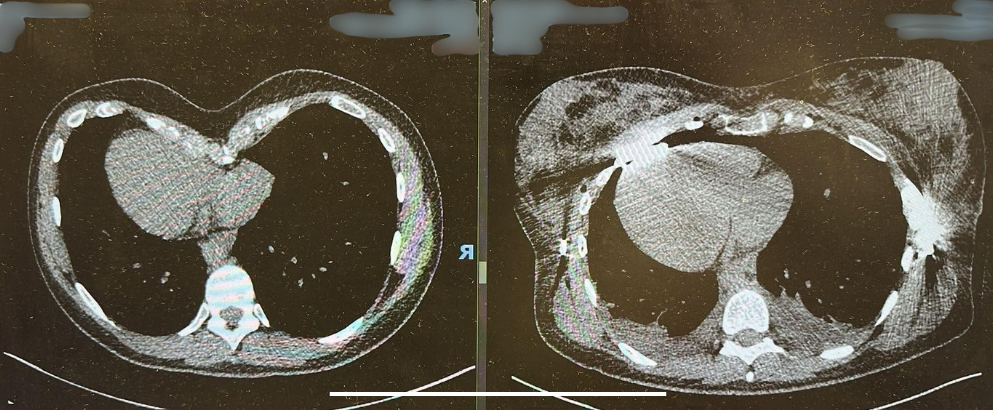
CT scan of a patient with pectus excavatum, before and after corrective surgery
-
An echocardiogram is an ultrasound scan of the heart that shows how the heart is functioning.
During an echocardiogram, the patient lies down on an exam bed whilst a sonographer moves a small probe across their chest. An image of the heart will appear on a screen beside you. The sonographer will take some measurements of the images, which will show how well your heart is functioning. Echocardiograms can take between 5-15 minutes to complete. During the scan, the sonographer might ask you to turn onto your side and take some deep breaths, so they can get the best image possible.
-
A cardiac MRI scan produces highly detailed images of the heart’s structure, valves and major blood vessels. If you have pectus excavatum, your consultant may request that you have a cardiac MRI to see whether the heart’s ability to function has been compromised by the deformed sternum.
MRI scanners are large and tunnel-shaped. They create detailed images by spinning magnets around the outside of the tunnel, where the patient lies. Before a cardiac MRI scan, a cannula will usually be placed in your arm. The patient lies down on a bed which moves in and out of the tunnel. The cannula in your arm will be connected to the scanner, which will inject dye into your arm during the scan, to help make the muscles of the heart more visible.
Before the scan starts, you might be given headphones to wear to dampen the noise and so that the radiographer can talk to you and give you instructions throughout the scan.
During the scan, the scanner will make a loud noise as the magnets spin around the outside of the tunnel.
The radiographer will speak to you through the headphones and ask you to take some deep breaths or hold your breath for a few seconds.
-
Creating a 3D scan of the chest is a modern technique that maps the shape of the sternum and helps visualise the severity of a pectus deformity.
These scans can be used in patients with pectus excavatum to measure severity and assist with the design of custom-made pectus implants.
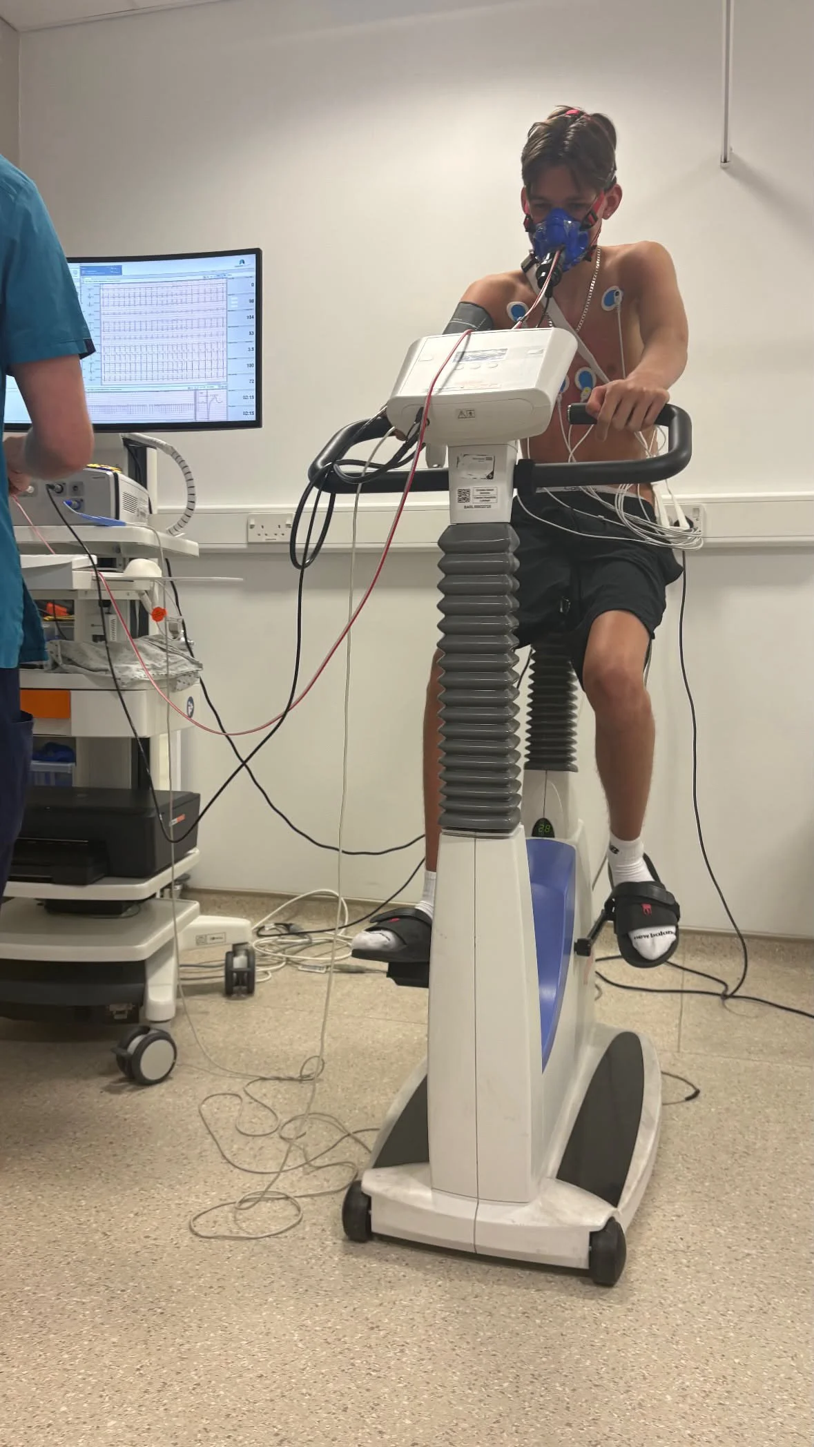
A patient with pectus excavatum having a cardiopulmonary exercise test (CPET)
3D scans taken of a patient with pectus excavatum, before and after the NUSS procedure